Basal cell carcinoma and squamous cell carcinoma are the two most common forms of skin cancer diagnosed in the UK. Between them, they account for the vast majority of skin cancer cases, and for both, surgical excision is frequently the treatment of choice.
Despite being mentioned in the same breath, they are different conditions with different behaviours, different risks, and sometimes different surgical approaches. This post explains what each one is and how excision surgery addresses them.
Basal cell carcinoma: the most common skin cancer in the UK
Basal cell carcinoma, or BCC, arises from the basal cells that sit at the deepest layer of the epidermis, the outer skin. It is the most frequently diagnosed skin cancer in the United Kingdom, with around 147,000 new cases recorded each year according to Cancer Research UK.
BCC grows slowly. It rarely spreads to other parts of the body, which is why the outlook following prompt treatment is very good. What it can do if left untreated is grow locally, invading surrounding tissue and becoming progressively harder to treat. The question that comes up often in consultations is what happens if a BCC is left for two years or more? The honest answer is that it will continue to grow and the surgical margins required to achieve clear removal will increase accordingly. Early treatment is always easier.
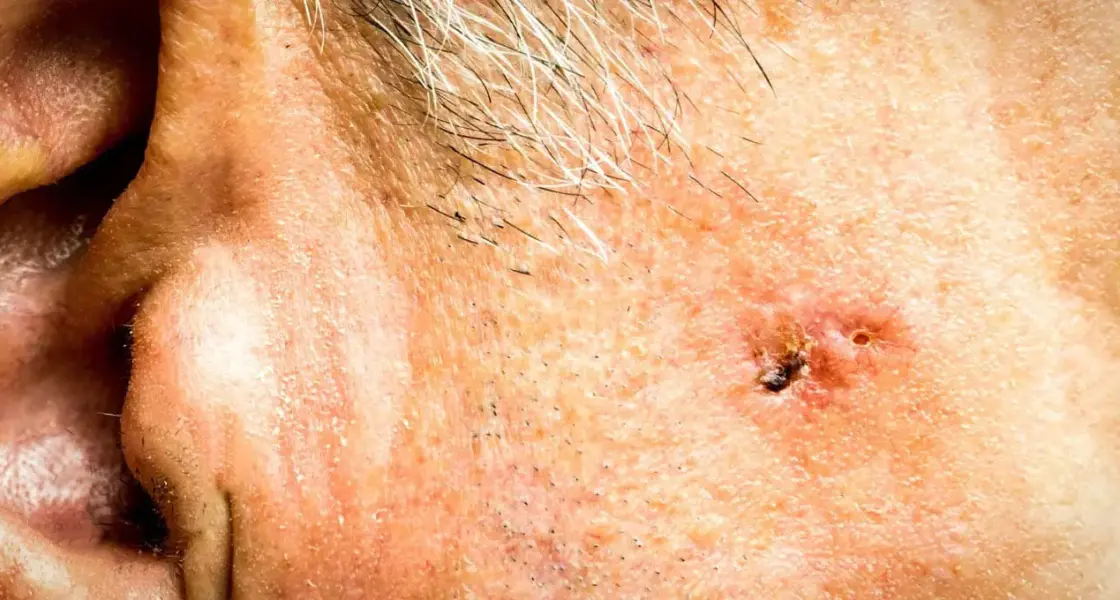
BCC most commonly appears on sun-exposed areas, particularly the face, scalp, ears, and neck. It often looks like a small, shiny or pearlescent lump, sometimes with a slightly rolled border. Some present as a flat, pink or pale patch. If you notice something that has been slowly changing over months and fits that description, it warrants assessment.
Squamous cell carcinoma: more aggressive, still very treatable
Squamous cell carcinoma, or SCC, arises from the squamous cells in the upper layers of the skin. It is the second most common skin cancer in the UK. SCC grows more quickly than BCC and carries a small but real risk of spreading to lymph nodes or other organs if not treated promptly, which is why early diagnosis and definitive treatment matter more here.
SCC often presents as a firm, red nodule or a flat lesion with a scaly, crusted surface. It can develop from pre-existing actinic keratoses, rough, scaly patches that form on chronically sun-damaged skin, or arise de novo. Like BCC, sun-exposed areas are the most common sites, though SCC can also occur on areas that have been subjected to chronic inflammation or previous radiation.
The prognosis for SCC treated at an early stage is excellent. Most patients who undergo surgical excision with clear margins are cured. The risk of recurrence or spread increases when tumours are larger, deeper, or located in higher-risk anatomical sites such as the lip or ear.
How surgical excision works for skin cancer
Surgical excision removes the tumour along with a margin of surrounding normal-appearing skin. That margin is deliberate; it accounts for the fact that cancer cells can extend beyond what is visible on the surface, and clear margins confirmed on histology are the goal.
The required margin varies by cancer type and tumour characteristics. For a straightforward BCC in a low-risk site, a margin of 3 – 4 mm is typically sufficient to achieve clearance. For SCC, or for larger or higher-risk BCCs, wider margins are used. The excised tissue is always sent for histological analysis (laboratory examination) to confirm that the margins are clear.
The procedure is performed under local anaesthetic and takes between 30 and 60 minutes depending on the size and location of the lesion. Most patients are surprised by how straightforward it is on the day. You are awake, comfortable, and go home the same day.
What happens if margins are not clear?
Occasionally, histology shows that cancer cells extend to the edge of the excised tissue, meaning further surgery may be needed to achieve complete clearance. This is not a complication; it is a planned contingency, and the decision to re-excise is made based on the pathology result rather than assumption. Your surgeon will discuss this with you if it applies.
Reconstruction after skin excision
For small lesions in favourable locations, the wound is closed directly with sutures and heals very well. For larger excisions, or those in anatomically complex areas like the face, a more involved closure may be needed, such as a local flap, using adjacent skin to reconstruct the area, or occasionally a skin graft for defects that cannot be closed primarily.
This is where the plastic surgical element of the work matters. Achieving clear oncological margins and an acceptable cosmetic result in the same procedure requires planning, and for lesions on visible areas like the nose, eyelid, or lip, the reconstructive approach is as important as the excision itself.
Recovery after skin cancer excision
Most patients return to normal daily activities within a week. The wound will be closed with sutures, either dissolvable or removable, depending on the site, and you will be given aftercare instructions from our professional staff covering wound care, activity restrictions, and signs to look out for.
Healing time varies by site and the complexity of the closure. A simple excision on the back will heal differently to a flap closure on the face. Scars mature over twelve to eighteen months, and the final appearance of the scar is generally much better than patients anticipate in the early weeks.
For anyone who has been told they have a BCC or SCC, or who has noticed a changing skin lesion that has not yet been assessed, a surgical consultation with Dr Abood is worth arranging promptly. You can find full details about the procedure on the surgical excision treatment page.
Book a consultation
Full details about surgical excision for skin cancer are on the treatment page at dr-abood.co.uk/treatments/surgical-excision/
Consultations are available at Dr Abood’s clinics in London and Cambridge. Book at dr-abood.co.uk/contact-us/
References
[1] NHS (2023). Basal cell carcinoma (BCC). nhs.uk/conditions/basal-cell-carcinoma
[2] NHS (2023). Squamous cell carcinoma (SCC). nhs.uk/conditions/squamous-cell-skin-cancer
[3] Cancer Research UK (2023). Basal cell carcinoma incidence statistics. cancerresearchuk.org
[4] British Association of Dermatologists (2021). BAD guidelines for the management of BCC. bad.org.uk
[5] NICE (2022). Skin cancer: recognition and referral. nice.org.uk/guidance/ng12
[6] Motley, R. et al. (2021). Multiprofessional guidelines for the management of the patient with primary cutaneous SCC. British Journal of Dermatology. 185(1): 18-33.
[7] Telfer, N.R. et al. (2008). Guidelines for the management of basal cell carcinoma. British Journal of Dermatology. 159(1): 35-48.